Tuesday 27 February 2018

observation of permanent slide for mitosis and meiosis


Observation of permanent slide for mitosis and meiosis
Aim: To observe the different stages of meiosis using permanent slides
Principle: Meiosis is a type of cell division in which the number of chromosomes is halved (from diploid to haploid) in the daughter cells, i.e., the gametes. The division is completed in two phases, meiosis I and meiosis II. Meiosis I is a reduction division in which the chromosomes of homologous pairs separate from each other. Meiosis II is equation division resulting in the formation of four daughter cells. Stages of meiosis can be observed in a cytological preparation of the cells of testis tubules or in the pollen mother cells of the anthers of flower buds.
Requirement: Permanent slides of meiosis and compound microscope
Procedure: Place the slide on the stage of the microscope and search for the dividing cells using lower magnification. When dividing cells are located observe them under higher magnification.
Observation:
 Various stages of meiosis were identified on the basis of the specific features present in the slide. A significant number of cells will be in the Interphase.

Leptotene:The nuclear membrane and nucleolus are not distinctly observable.
Zygotene: This stage is characterised by the pairing of the homologous chromosomes, which can be seen as paired chromatin threads (bivalents)
Pachytene: The chromatin threads get condensed and appear shortened and thick. Pairs of homologous chromosomes and tetrad can be seen.
Diplotene: The homologous chromosomes show distinct separation from each other except at few regions where attachments are seen. These are called chiasmata where crossing over occurs.
Diakinesis: Nucleus division can be seen.
Metaphase I: At this stage, the number of bivalents can be counted. Chiasmata may still be seen in a few bivalents.
Anaphase I: This stage can be identified the presence of two chromatids in each chromosome.
Telophase I: The chromosomes present at the two poles appear decondensed and form two distinct nuclei.
Prophase II: (i) Distinct thread- like chromatin fibers or rod- shaped chromosome is seen.
Metaphase II: In the metaphase I of meiosis, a few chiasmata are observed, where as no chiasmata are observed during metaphase II.
Anaphase II: The two chromatids of each chromosome after separation appear to lie at the two poles of the cell
Telophase II: The separated chromosomes appear de condensed and form nuclei.
 STUDY OF THE STAGES OF MITOSIS FROM PERMANENT SLIDES
Aim: To observe the different stages of mitosis using permanent slides
Requirement: Permanent slides of mitosis and compound microscope
Procedure: The permanent slide was placed on the stage of compound microscope and observed the stages of mitosis.
Observations:
Various stages of meiosis were identified on the basis of the specific features present in the slide.
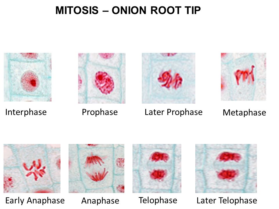
1. Prophase: In this slide some chromosomes are seen. The chromosomes are long and scattered. No spindle fiber is seen. Therefore the stage is Prophase of Mitosis.
2. Metaphase: Some chromosomes are seen in this slide. Spindle apparatus is seen here. The chromosomes are situated on the equatorial zone. The chromosomes are divided into chromatids. Therefore this is the Metaphase of Mitosis.
3. Anaphase: In this slide two sets of chromosomes are seen. Two sets are present near the two poles. Therefore it is the Anaphase of Mitosis.
4. Telophase: In this slide two sets of chromosomes are seen. Two sets of chromosomes are present at two poles. No spindle apparatus is seen. Nuclear membrane is present surrounding the chromosomes in each pole. Therefore it is the Telophase of Mitosis.
Note:some content obtained from web source.

Thursday 15 February 2018

bacterial cell count by counting chamber


Bacterial Cell count by counting chamber
 AIM: To count the number of microorganisms in a given sample by using counting chamber.
 INTRODUCTION: For unicellular microorganisms, such as bacteria, the reproduction of the cell reproduces the entire organism. Therefore, microbial growth is essentially synonymous with microbial reproduction. To determine rates of microbial growth and death, it is necessary to enumerate microorganisms, that is, to determine their numbers. It is also often essential to determine the number of microorganisms in a given sample. For example, the ability to determine the safety of many foods and drugs depends on knowing the levels of microorganisms in those products. A variety of methods has been developed for the enumeration of microbes.  Direct Count of Cells by counting chamber: Direct microscopic counts are performed by spreading a measured volume of sample over a known area of a slide, counting representative microscopic fields, and relating the averages back to the appropriate volume-area factors. Specially constructed counting chambers has the ability to count a defined area and convert the numbers observed directly to volume makes the direct enumeration procedure relatively easy. Direct counting procedures are rapid but have the disadvantage that they do not discriminate between living and dead cells. This method is used to assess the sanitation level of a food product and in performing blood cell counts in hematology and microbial cells in microbiology.
Materials required:
Direct Count Using a Counting Chamber, Bacterial suspension, lab Counting chamber, Pipettes.
Calculating cell count
The total number of cells per microliter of sample can be calculated from the number of cell counted and area counted. This is because the ruled areas of the chamber contain an exact volume of diluted sample. Since only a small volume of diluted sample is counted, a general formula must be used to convert the count into the number of cells/microliter.
The dilution factor used in the formula is determined by the blood dilution used in the cell count. The depth used in the formula is always 0.1. The area counted will vary for each type of cell count and is calculated using the dimensions of the ruled area.
The smallest square has an area of 0.0025 mm2 therefore each main square has an area of 0.04 mm2 (0.0025 x 16 = 0.04).

The depth of the chamber is 0.100 mm (space between the glass slide and cover slip) then the volume is calculated as:
(0.04) x (0.100) = 0.004 mm3 = 0.004 μl
To calculate the amount of cells in 1μl a rule of three is applied. If we have X amount of cells in 0.004μl, how many cells are in 1μl?:
# cells -------------0.004 μl
? --------------1μl
Then:
Cells in 1μl = (number of cells in a main square)(1μl) /0.004
Cells in 1 ml = (cells in 1μl) x (1000)

Example:
Lets calculate total WBC count by using Neubauer counting chamber.
Number of cells counted = N = 150 (suppose)
Area Counted = 1 mm2 x 4 = 4 mm2 (area of four large corner squares)
Depth = 1/10 mm
Dilution = 1:20
Hence WBC/Cubic mm of Whole Blood = N x 50 = 150 x 50 = 7,500

 Procedure:
 1. Clean a counting chamber with lens paper and then place it on the microscope stage.
2. Using the 40X objective find the ruled area and arrangements of larger squares and their small square subdivisions.
 3. Shake the sample suspension to distribute the cells evenly. Take out the counting chamber without changing the focus on the 40X objective. Place a coverslip over the counting chamber.
4. Using a transfer pipette, transfer some of the suspension to the groove of the counting chamber to fill the chamber by capillary action.
5. Carefully place the counting chamber back onto the microscopic stage and observe the cells under 40X.
6. Count the number of cells in at least 50 of the small squares. If cells fall on a line, include in your count those on the top and left-hand lines and exclude those on the bottom and right-hand lines.
7. Calculate the average number of cells per Small Square. Then calculate the number of per ml by dividing the average number of cells per Small Square by the volume of each small square which is 0.00025 µl. If diluted the sample, also multiply the results by the dilution factor to determine the concentration of cells in the original sample. Record the calculations and results.
Result:
Direct Counts Using A Counting Chamber
Total number of yeast cells counted __________________
Average number of cells per counting squares _____________
Volume associated with each counting square __________________
Dilution factor (if any)_____________
Concentration of yeasts in original sample____________________

Thursday 1 February 2018

MICROMETRY


MICROMETRY
AIM: To measure the size of the given microorganism by using micrometry.
PRINCIPLE: Measuring the size of microorganism using a microscope is called micrometry. All measurements of length are based on a comparison of the object under scrutiny with known dimensions, or with a standardized, calibrated scale. In order to determine the length or width of a microbe, for example, a ruler or measuring tape is placed in contact with the board and the dimensions are noted by direct comparison to the graduated numerical markings on the ruler. This basic principle is applicable to the measurement of specimens observed in the microscope, is often not possible with a compound microscope to place a ruler in direct contact with the specimen. Alternative mechanisms for performing measurements at high magnifications in compound optical microscopy must be employed, and the most common of these is the application of eyepiece reticles in combination with stage micrometers. A majority of measurements made with compound microscopes fall into the size range of 0.2 micrometers to 25 millimeters. Horizontal distances below 0.2 micrometers are beneath the resolving power of the microscope, and lengths larger than the field of view of an eyepiece are usually measured with a microscope.
MATERIALS REQUIRED
Light microscope, ocular and stage micrometer, slides, bacterial cell culture

PROCEDURE
1. The ocular micrometer is placed on the circular shelf inside the eyepiece of the microscope.
2. Place the stage micrometer on the stage of a microscope and focus the graduations using low power objectives. The graduations on stage micrometer are spaced 0.01mm (10μm) apart.
3. Superimpose the two scales and record the number of ocular division coinciding exactly with the number of divisions of the stage micrometer.
4. The calibration factor or the least count of ocular micrometer is calculated
5. Now remove the stage micrometer from the stage and place the slide having cell preparation under low power magnification.
6. Position the cell being observed in such a way that the ocular micrometer is able to measure the diameter of a cell or the length/diameter of a cell component in arbitrary units.
7. Similarly for high power objective the ocular micrometer calibration has to be done again following the same procedure and then cell diameter is can be measured focusing the cell in high magnification.
8. Measure a rod-shaped bacterium, a coccus, a yeast cell, a protozoan, and a human red blood cell.  Record the results. 
Detection of calibration factor as follows:
If 13 ocular divisions coincide with 2 divisions (2X10μm=20μm) of stage micrometer
Then 1 ocular division =         20μm
-------                           = 1.54μm
13 divisions
Calculate the size as shown below:
If the diameter of a cell is occupying 5 divisions of ocular, the diameter of the cell will be:
 5 divisions X 1.54μm = 7.7μm

OBSERVATION:
The size of the given organism was measured with micrometry.
RESULT:
The size of the given organism was tabulated
S.No
Type of organism
Objective lens magnification
Calibration value
Size(mm)
1.
Bacteria/yeast/algae
10X


40X


100X


 https://onedrive.live.com/edit.aspx?resid=6BF90612664526FF!520&cid=6bf90612664526ff&CT=1698476756796&OR=ItemsView https://onedr...